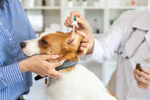
Let’s learn more about otitis externa in dogs, along with its symptoms, diagnosis, and treatment.
Otitis external (outer ear infection) is prevalent in dogs. Although ear infections can occur in any breed, it mainly affects those with large, hairy, or floppy ears as well as those prone to allergies, such as Miniature Poodles, Cocker Spaniels, or English Bulldogs. Let’s learn more about otitis external, along with its symptoms, diagnosis, and treatment.
Dog Ear Anatomy
The anatomy of a dog’s ear consists of the ear flap, ear canal, inner ear, eardrum, and middle ear. The outer ear includes the pinna (the cartilage part of the ear that is below skin, fur, or hair) and the ear canal. The pinna captures the sound waves and releases them through the dog’s ear canal to the eardrum. The middle ear includes 2 muscles, an oval window, and the eustachian tube. The eustachian tube is a small tube that ties the middle ear with the back of the dog’s nose, allowing air to enter the middle ear. Lastly, the inner ear includes the cochlea (the organ of hearing) and the vestibular system (the organ for balance).
In people, a short, straight canal connects all the parts of the ear. In dogs, the canal is much deeper and bends as it goes into the middle ear. This anatomy creates an environment that promotes bacterial and yeast growth, making dogs naturally more prone to ear infections.
What are the Symptoms of an Ear Infection in Dogs?
Generally, ear infections can be painful. Many dogs scratch their ears and shake their head to relieve the discomfort. In addition, their ears will become red and swollen, forming an odd scent. Your dog’s ears may also seem dirty with dark debris or with discharge coming from your dog’s ears. The ears might appear crusty in chronic cases, and the ear canals often become narrowed due to chronic inflammation.
Diagnosis of Otitis Externa in Dogs
Nearly all ear infections can be successfully handled with a proper diagnosis and treatment. However, the outcome will worsen if an underlying cause is unidentified and untreated. There are multiple kinds of bacteria and at least one kind of fungus that commonly produces ear infections. It’s impossible to know which medication to use without knowing the specific type of infection. So, multiple examinations will have to occur before the result is successful.
First, your dog’s ear canal is examined with an otoscope, an instrument that zooms into the ear with a built-in light. This vet examination allows us to determine whether the eardrum is intact or if there is any abnormal material in the ear canal, such as a tumor, foreign body, or a polyp. It may also be necessary to sedate your dog for a thorough exam if he or she is in extreme pain and refuses to allow the examination.
Secondly, we will determine the cause of the ear infection by examining a sample of the ear canal under a microscope. Microscopic examination is vital in assisting us in selecting the proper medication to treat your dog’s inflamed ear canal. The results of the microscopic examination typically determine the diagnosis and treatment. In more severe cases, a culture may be necessary to identify the specific organism causing the infection.
Furthermore, identifying any potential underlying disease is crucial to the patient’s evaluation. Many dogs with recurrent or chronic ear infections have low thyroid function or allergies. A vet must diagnose or treat the potential underlying disease, or the pet will continue to experience chronic ear issues.
Treatment
For most cases of otitis externa, we will clean your dog’s ears to remove any debris and then instill an ear medication in the hospital or send you home with medication to administer for an extended course of treatment. More severe cases might require oral medication or even an ear flush with our digital otoscope. An ear flush allows the doctor to better visualize the deeper parts of the canal and remove excessive or stubborn debris. It allows us to further explore the canal for foreign material, tumors, or polyps if your dog is experiencing recurring issues.
Untreated or chronic otitis externa can lead to the dog’s ear canal closing – stenosis or hyperplasia. It’s difficult for medications to enter the horizontal canal if the dog’s ear canal is swollen. Anti-inflammatory medications can sometimes shrink the swollen tissues and open the ear canal in some cases. In other cases of hyperplasia, surgery is necessary.
There are multiple surgical procedures that can alleviate chronic and severe ear issues. A total ear ablation and bulla osteotomy (TECA) is a common procedure for dogs experiencing these issues. This surgery involved the complete removal of the ear canal and tympanic bulla, or middle ear. This surgery ultimately removes the infected and abnormal tissue, reducing the cause of the inflammation that is painful to your dog.
Here at Mount Carmel Animal Hospital, We’ll Treat Your Pets Like Family!
Mount Carmel Animal Hospital has been serving the Northern Baltimore/Southern York community for over 30 years and is proud to be an independently operated, small animal practice committed to excellence in veterinary medicine and client service. From grooming to wellness services, along with Canine Life Skills Training Courses, and surgical procedures, we have the expertise that will best serve the needs of you and your pet. Contact us at 410-343-0200 and follow us on Facebook!
