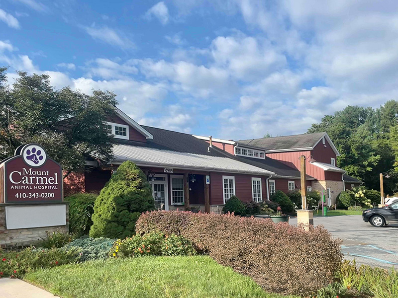
Mount Carmel Animal Hospital
Helping pets and people for over 30 years
Welcome New Clients
Now Offering CT Scans
Refill RX & Food

About Us
Trusted Veterinary Care for Your Pets at Mount Carmel Animal Hospital
Mount Carmel Animal Hospital (MCAH) has been serving the Northern Baltimore – Southern York community and under the same ownership for over 30 years. Together, Dr. Paul Fox and Dr. Matt McGee have worked to build MCAH into one of the premier practices in our region. We are proud to be an independently owned and operated small animal practice. We are committed to excellence in veterinary medicine and client service.
Veterinary Services
Complete Veterinary Services in Northern Baltimore County
At Mount Carmel Animal Hospital in Monkton, MD, we provide comprehensive veterinary services designed to meet the diverse needs of pets at every stage of life. Our wellness care focuses on preventive measures to keep your pets healthy, while our medical treatments address various health concerns with precision and care. We utilize advanced diagnostics and internal medicine expertise to accurately identify and manage complex conditions. Our surgical services and rehabilitation therapy ensure optimal recovery, while home care support and wellness care provide convenience and peace of mind. Additional offerings such as microchipping services and medical acupuncture further enhance your pet’s well-being, making us a trusted partner in their lifelong health journey.

Pet Wellness Care

Pet Dentistry

Pet Surgical Services

Pet Advanced Diagnostics
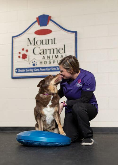
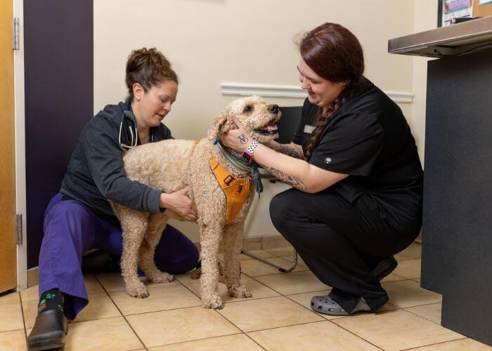
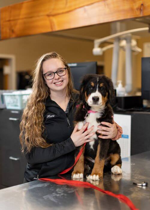
Meet Our Veterinary Team
Dedicated, compassionate, and highly skilled, our veterinary team is committed to providing exceptional care for your pets. With expertise spanning wellness, advanced diagnostics, and specialized treatments, we work together to ensure every patient receives personalized and attentive care.


